


Preserving Prehistoric Snacks: 3D Scanning a Unique Pterosaur Fossil
Updated: Jul 30
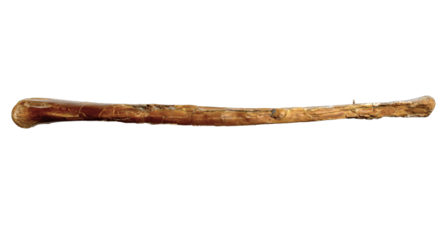
In the world of 3D scanning, Rapid3D is lucky enough to work with a lot of fascinating objects. Recently, our team had the privilege of working with the Royal Tyrrell Museum to 3D scan an extraordinarily rare piece of natural history: a pterosaur tibia.

Image Credit: Royal Tyrrell Museum of Palaeontology
Art by Julius Csotonyi
There are a few reasons why this fossil is so special and interesting.The tibia fossil, which is the lower leg bone of an azhdarchid pterosaur, is the most complete tibial specimen found in Canada and was excavated in Dinosaur Provincial Park in the 1990s. Pterosaur fossils are rare because the animal’s bones were thin and hollow. The distal end (near the ankle) of this tibia specimen has small tooth marks, confirmed by the fact the fossil contains a tooth embedded right in the bone! This is an undeniably neat snapshot of a mealtime interaction from millions of years ago. The tooth has been identified as coming from a dromoaeosaur or raptor, and it is theorized that the raptor was scavenging on the already-dead pterosaur since there is no sign of bone healing around the tooth. (Source: Everything Dinosaur).

To read more about this specimen, visit the Royal Tyrrell Museum’s online collections database. The object number is TMP1992.083.0002
To preserve this remarkable specimen and ensure it can be studied and shared without risking damage to the original, we needed to capture both its detailed geometry and its visual texture appearance. We used two of FARO Creaform’s high accuracy 3D scanners to make this challenge an easy one to accomplish.
The Challenge: Capturing Geometry and Color
This pterosaur tibia presented a unique challenge. The embedded tooth is quite small, meaning the scan needed to capture tiny variations in geometry and surface detail to be of any use to paleontologists and to ensure an accurate duplication. At the same time, the surface colour is an important feature to preserve as well.
To get the best of both worlds, we utilized two scanners in our approach, combining two cutting-edge tools to create the ultimate digital twin.
1. Capturing the Details with the HandySCAN EVO Elite
To capture the high resolution detail of the bone and the embedded tooth, we utilized the HandySCAN 3D EVO Elite. We needed the High-Resolution Accuracy, which allowed us to capture the finest details of the bone and tooth surface geometry. Because 3D scanning is fully optical, there was no need to touch the fossil or place targets on the tibia itself. We were able to use 6mm positioning targets and 360 target balls placed around the fossil for positioning.

2. Bringing It to Life with the Go!SCAN SPARK
While the HandySCAN mapped the exact physical dimensions at high resolutions, we needed to capture the authentic look of the fossil. For this, we used the Go!SCAN SPARK.
● Realistic Texture: This white-light scanner is designed to quickly map colour and texture over 3D geometry.
● Seamless Integration: By combining the SPARK’s colour data with the EVO Elite’s geometric accuracy, we created a complete 3D model that looks and measures exactly like the real thing sitting on the lab table.

From Digital Twin to Physical Replica
So, where does this incredible digital data go from here?
The final, fully textured high-resolution 3D scan has been sent to the Royal Ontario Museum (ROM). Using the watertight Rapid3D scan data, the ROM will 3D print a physical replica of the pterosaur tibia.
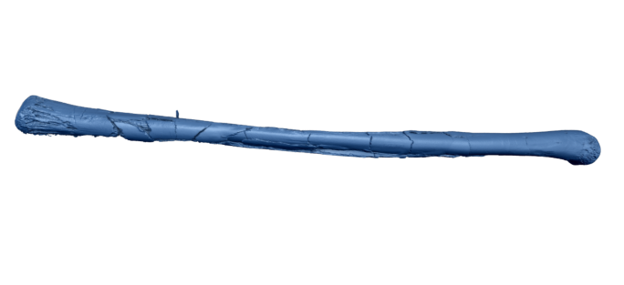

This means that while the fragile original remains safely preserved at the Royal Tyrrell, researchers, educators, and museum visitors at the ROM will be able to handle, study, and marvel at a perfect physical copy of this ancient dinnertime encounter.
Projects like this highlight exactly why we love what we do. By merging paleontology with state-of-the-art 3D metrology, we aren't just measuring objects, we are helping museums preserve our planet's history and making the ancient world more accessible to everyone!


What's up guys, I was listening to a podcast about mobile gaming while commuting on the train. I’d blown my budget for the month on a series of awful dice games. I felt completely defeated and was plotting how to explain my missing funds. The podcast host casually dropped the name, so I opened Mr Beast Casino on my mobile browser. The mobile optimization is flawless, which is a huge plus for anyone navigating across EN with spotty internet. I played a simple Italian-flair slot while riding home. I matched enough symbols out of sheer dumb luck. The win was large enough to completely erase my previous failures and put me firmly in the green. I really appreciated the seamless mobile…
I usually avoid making a decision after reading just one review because it's too easy to miss the bigger picture. Instead, I check several discussion threads where people compare their experiences over time. That approach helped me filter out a lot of noise. Treasure Cove Casino was one of the few names that kept appearing in different conversations, and the feedback was consistently measured. Players seemed to appreciate the overall experience without describing it as something extraordinary, which actually made the recommendations feel more believable.